Radiology in dentistry means radiography or X ray. Dentists mainly depend on radiographs to know the structures hidden like cavities, bone loss and masses which are malignant and benign. Both tooth decay and periodontal disease can be missed during a clinical examination, hence, radiographic evaluation of the dental and periodontal tissues is a crucial part of the comprehensive oral examination.
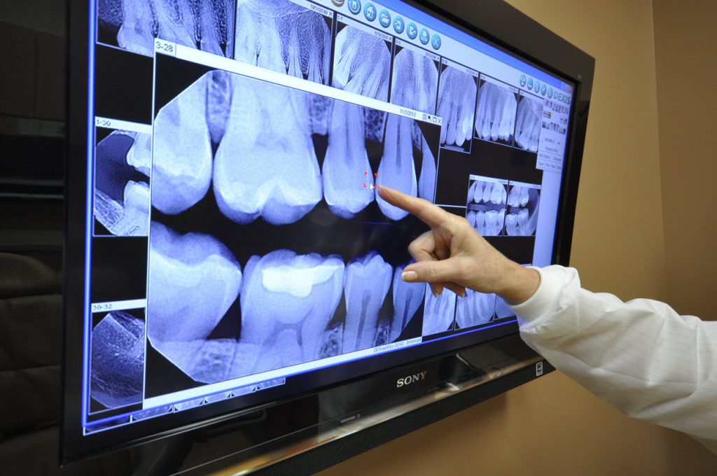
Digital radiography-The digital radiography is very similar to the traditional dental X-rays that use film. In digital radiography, dentist inserts a sensor into your mouth to capture images of the teeth. This is the only similarity between traditional and digital dental X-rays. Here a digital sensor is used to scan the teeth instead of the film. It is highly secure and quality scanning.
There are two types of Radiography
CR uses a photostimulable phosphor (PSP) plate to capture images. A laser scanner then scans the plate causing the stored energy (image) to release and subsequently captured to create the digital image.
DR uses a charge-coupled device (CCD) or a complementary metal oxide semiconductor (CMOS) sensor. Both these sensors are attached to a wire which is used to transfer the image from the sensor to a computer. The imaging of the CCD and CMOS sensors are almost similar.
Digital radiography requires less radiation than film radiography. The patient radiation dose for CR is in the range of 100 to 125 mrad per image and the dose for DR is in the range of 50 to 75 mrad per image. It is good to reduce the exposure time by at least one half when changing from D-speed film to CR. The exposure time must be less than 0.2 seconds (2/10 seconds; 200 milliseconds) for digital imaging. If the exposure time increases more than 0.2 seconds, it is considered as over dosage for diagnostic images.
There are two views
Authored By Dr Sanjay N - Orthodontics & Dentofacial Orthopaedics, Bangalore